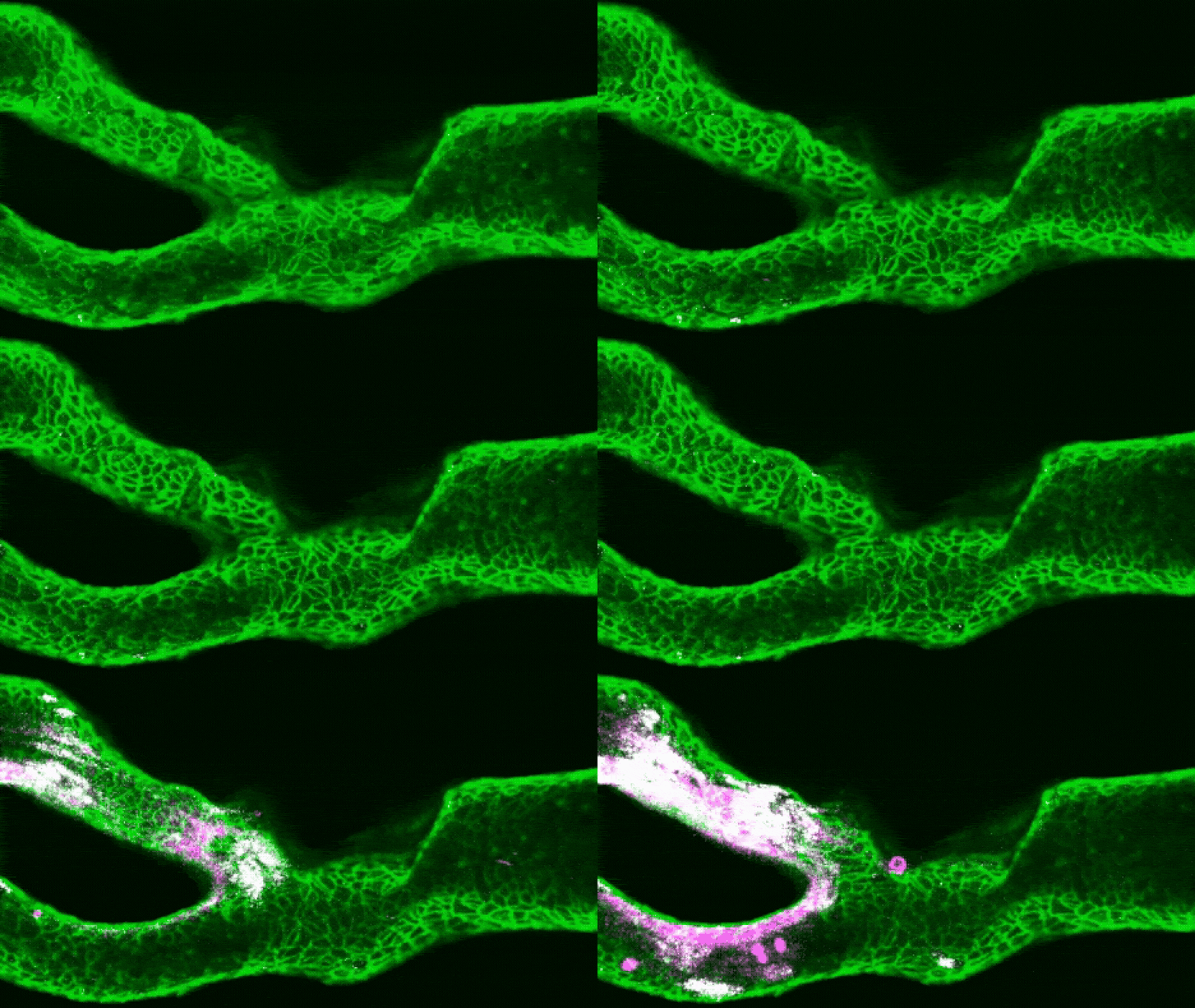
Researchers have pioneered a way to create miniaturized copies of a person's carotid artery that could help doctors predict and manage the patient's stroke risk, a new study suggests.
In the research, published in July in the journal Cell Biomaterials, scientists used these "arteries-on-a-chip" to monitor how real blood flows through a patient's carotid arteries, which carry blood to the brain, face and neck. This could enable doctors to identify not just how and what type of clots form in that specific patient, but also determine which medications would be most effective in dealing with the blockage.
While the technique is currently a proof-of-concept, someday, it could help medical practitioners to better tailor treatments for each patient.
"I see this approach being most relevant to difficult clinical cases," said study first author Charles Zhao, a doctoral student at the University of Sydney. "For example, patients who have experienced recurrent events despite treatment, or cases where clinicians have several possible treatment strategies but limited functional information to distinguish between them." Zhao told Live Science in an email.
Ischemic stroke is one of the leading causes of death worldwide, second only to heart disease. The medical emergency is most often caused by a loose blood clot blocking an artery in the brain, starving the cells of oxygen. Obesity, high blood pressure and high cholesterol are all important risk factors for ischemic stroke, and roughly 1 in 5 of these events results from atherosclerosis, or fatty plaque buildup in the arteries.
"When the inner lining of an artery is damaged, material underneath the cells, including collagen, becomes exposed to the blood," Zhao said. A bloodborne protein called von Willebrand factor (VWF) grabs hold of platelets from the flowing blood. Those platelets then stick together and recruit more platelets, building a clot, he said.
But forming a clot is only half the story; it's what this mass does next that determines the risk to the patient.
In some cases, the growing ball of clotted blood will stay firmly rooted to the artery wall, slightly impeding blood flow but not posing any immediate danger. However, if tiny fragments break off this static clot, they can travel toward the brain, where they risk blocking smaller vessels and thus causing a stroke.
"Clinically, we are very good at imaging how narrow an artery is," Zhao said, "but narrowing alone does not tell us exactly how a clot will behave."
This is where artery-on-a-chip models come in, he said. Rather than relying purely on medical scans, the team's model recreates the exact shape and structure of an individual patient's blood vessels using 3D printing. To create the models, they first use a patient’s existing CT scans to 3D print a plastic replica of their carotid artery, including any narrowing caused by atherosclerosis. Next, the inside of this plastic structure is coated with collagen, and then cells that line the carotid artery get layered on top. Blood is passed through the replica artery, mimicking the speed and pressure of blood flow in the body.
Study co-author Zihao Wang holding an assembled 3D-printed blood vessel device. (Image credit: University of Sydney/Fiona Wolf)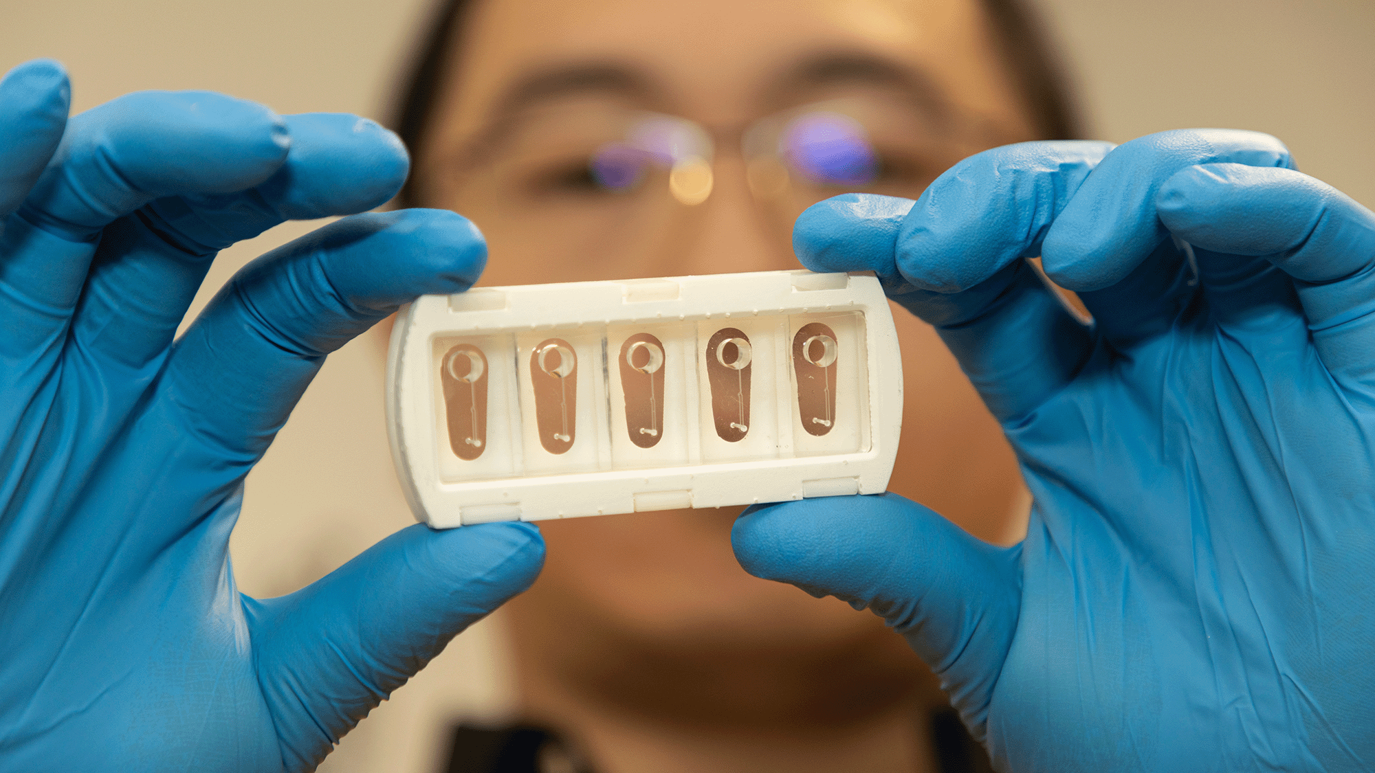
"Then [we] use a very small laser injury to expose the collagen underneath," Zhao explained. "We can then flow human blood through the artery and watch thrombosis happen under a microscope." Thrombosis is the formation of a blood clot in a blood vessel.
The researchers created these models for six patients with different types of arterial damage and used various calculations to understand the impacts of changes in blood flow through the structures.
By combining these calculations with the patient-specific artery structures, as well as tests using real blood flow, the researchers built up a far more accurate picture of how clots formed and shed than medical images alone could capture. This revealed striking differences between seemingly equivalent patients.
Related stories- Scientists infected a 'vagina on a chip' with gonorrhea — then cured it with a new antibiotic found by AI
- Scientists developing new 'heart-on-a-chip'
- New pocket-size model of ALS 'breathes and flows like human tissue'
"We found that two arteries with apparently similar disease can behave very differently because their three-dimensional shapes create different patterns of blood flow," Zhao said. These local conditions within the artery are important in determining whether a clot grows stably or becomes prone to shedding fragments, he added.
These functional differences are particularly important when thinking about how best to reduce a person's stroke risk. Some treatments prevent platelets clumping together into clots. Others prevent platelets from sticking to the artery walls, while still others inhibit the key clot-forming protein VWF. The most effective medication for a given patient will therefore depend on how their clot forms and grows, making these new physical models a valuable complement to traditional imaging, Zhao said.
The team now hopes to expand this research further with the ultimate goal of establishing artery-on-a-chip models as an additional diagnostic step between initial medical imaging and treatment selection for patients.
This article is for informational purposes only and is not meant to offer medical advice.
What do you know about the body's hardest-working muscle? Find out with our heart quiz!